I. Application
This machine is applied to abdominal, urology, obstetrics and gynecology, pediatrics, neonatology, superficial structure, small organs, musculoskeletal, heart and so on.
It is applied to cow, horse, sheep, swine, cat, dog.
Inspection in the process of embryo transfer.
Recognition of false pregnancy of pig, horse etc.
Diagnosis of diseases on reproductive systems like uterus, ovaries.
Diagnosis of pregnancy, fetal development, fetal vitality and gender identification.
Diagnosis of all kinds of animal mammary tumor, abscess, hematoma.
II. Features
1. Display mode: B, B + B, 4B, B + M, M.
2. Measurement, distance, circumference, area, volume, heart rate, pregnancy week (BP, GS, CRL, HC, AC, EDD, AFI) and the expected date of birth, fetal weight display, etc.,
3. Electronic focus: four electronic focus
4. Body mark: ≥ 90 kinds
5. Image mirroring: up and down, left and right, black and white , in any mode can be changed and operated.
6. Images can be 0 °, 90 °, 180 °, 270 °, 360 ° rotation
7. Through the trackball cursor to select the function package and measurement methods
8. Character display: date, time, name, sex, age, doctor, hospital, comment (full screen character editing) (can be mixed with letters, numbers, punctuation, arrows)
9. Cine loop: ≥ 512 frames, can be continuous to play or view by line.
10. Permanent storage: built-in 8G memory, ≥ 4000 images, support U disk storage (can be read)
11. Puncture guidance function;
12. Four kinds of frame correlation, with independent keys, can be adjustable, can be adjusted, you can also change by trackball cursor;
13. 6 kinds of line correlation, with independent buttons, can be adjustable, recycled visual adjustment, you can also change the trackball cursor.
14. 8 kinds of gamma correction, with independent buttons, can be adjustable, recycled visual adjustment, you can also change the trackball cursor.
15. Edge enhancement, 0-3, 4 kinds of adjustment, with independent keys, can be adjustable visually, can be adjusted cyclically, you can also change by trackball cursor;
16. Scanning range, 0-3, 4 kinds, with independent keys, adjustable, can be adjusted cyclically, you can also change by the trackball cursor;
17. Interface: RS-232 , VGA , VIDEO , 2 USB ports, DICOM
III. Specifications
1. Main unit system:
a) Digital beamforming
b) Real-time dynamic aperture imaging
c) real-time dynamic sound velocity trace
d) 8-segment digital TGC
e) Tissue harmonic imaging unit
f) Dynamic digital filtering
g) image enhancement, row correlation, frame correlation, point correlation, linear interpolation and other image processing technology
2. probe interface
IV. Function introduction
|
PARAMETER |
SPECIFICATION |
|
Display mode |
B, B + B, 4B, B + M, M. |
|
measurement, distance, circumference, area, volume, heart rate, pregnancy week (BP, GS, CRL, HC, AC, EDD, AFI) and the expected date of birth, fetal weight display, etc |
|
|
electronic focus |
four electronic focus |
|
body mark |
≥ 90 kinds |
|
Image mirroring |
up and down, left and right, black and white , in any mode can be changed and operated. |
|
images can be 0 °, 90 °, 180 °, 270 °, 360 ° rotation |
|
|
through the trackball cursor to select the function package and measurement methods |
|
|
Character display |
Character display 9. cine loop: ≥ 512 frames, can be continuous to play or view by line. |
|
permanent storage |
built-in 8G memory, ≥ 4000 images, support U disk storage (can be read) |
|
puncture guidance function; |
|
|
0-3, four kinds of frame correlation, with independent keys, can be adjustable, can be adjusted, you can also change by trackball cursor; |
|
|
6 kinds of line correlation, with independent buttons, can be adjustable, recycled visual adjustment, you can also change the trackball cursor. |
|
|
0-7, 8 kinds of gamma correction, with independent buttons, can be adjustable, recycled visual adjustment, you can also change the trackball cursor. |
|
|
edge enhancement, 0-3, 4 kinds of adjustment, with independent keys, can be adjustable visually, can be adjusted cyclically, you can also change by trackball cursor; |
|
|
scanning range, 0-3, 4 kinds, with independent keys, adjustable, can be adjusted cyclically, you can also change by the trackball cursor; |
|
|
scanning range, 0-3, 4 kinds, with independent keys, adjustable, can be adjusted cyclically, you can also change by the trackball cursor; |
|
|
Interface: |
RS-232, VGA, VIDEO, 2 USB ports, DICOM |
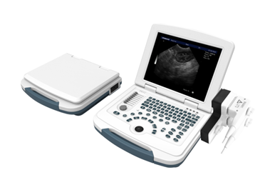
V. Product configuration
Main unit:
Main unit, power adapter PH80, 3 × 0.75MM power cord
12.1 inch medical LED display, 10400mA lithium battery
Gross Weight: 9.5KG
Manual
Warranty Card Certificate
Packaging plastic bags
Desiccant
Paper box
3.5MHz 80-element convex probe (optional)
6.5MHz 80-element micro-convex probe (optional)
7.5MHz 80-element rectal probe (optional)
7.5MHz 80-element high-frequency linear array probe (optional)
6.5MHz 80-element rectal probe (optional)
7.5MHz 80-element back fat probe (optional)
Frequency range:
3.5MHz: 2.0MHz, 2.5MHz, 3.5MHz, 4.0MHz, 5.0MHz
5.0 MHz, 4.0 MHz, 4.5 MHz, 5.0 MHz, 6.5 MHz, 7.0 MHz
6.5 MHz: 5.0 MHz, 5.5 MHz, 6.5 MHz, 7.5 MHz, 8.5 MHz
7.5 MHz: 5.5 MHz, 6.5 MHz, 7.0 MHz, 7.5 MHz, 9.0 MHz
VI. Technical Specifications
|
PARAMETER |
SPECIFICATION |
|
gray scale |
256 level |
|
Grayscale |
7 kinds, can be adjustable |
|
Dynamic range |
0-135dB |
|
Resolution |
horizontal ≤ 2mm; longitudinal ≤ 1mm |
|
Focus points |
1-4 focus optional, adjustable focus position continuously |
|
STC Gain Adjustment |
8 segments |
|
Tissue Harmonics |
0-1MHZ |
|
Maximum scan depth |
3.5mhz ≥ 307mm Visual adjustable 5.0MHz: 62-180mm 6.5MHz: 62-139mm 7.5MHz: 35-104mm |
|
Zoom function |
Overall zoom |
|
Image level adjustment |
0-1 |
|
black and white flip |
0-1 |
|
depth adjustment |
16 level |
|
Gain |
0 ~ 100 adjustable. |
|
scanning speed |
0-7 |
|
M pseudo color |
0-7 |
|
Blind zone |
≤ 4 |
|
Geometric accuracy |
horizontal ≤ 5%, vertical ≤ 5% |
|
Resolution |
lateral ≤ 2mm, axial ≤ 1mm |


